
Does medical imaging reconstruction technology cut dose?




Does Medical Imaging Reconstruction Technology Cut Dose?
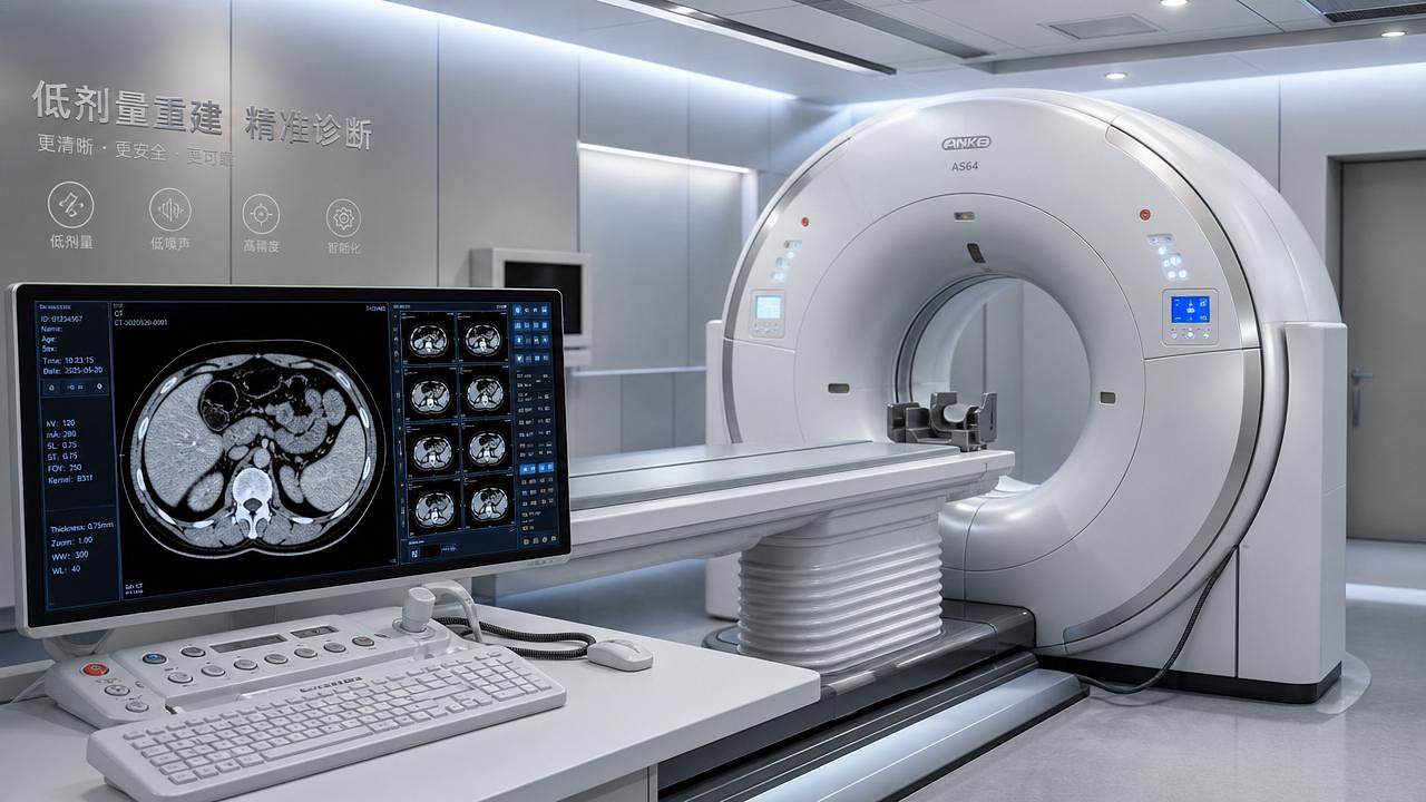
Does medical imaging reconstruction technology truly cut dose without compromising diagnostic confidence?
The answer depends on noise control, spatial resolution, artifact reduction, and clinical validation across CT, MRI, and photon-counting platforms.
As dose mandates tighten, reconstruction performance is becoming a strategic metric for safety, workflow, and precision diagnosis.
Dose Reduction Is Moving From Protocol Tuning To Algorithmic Evidence
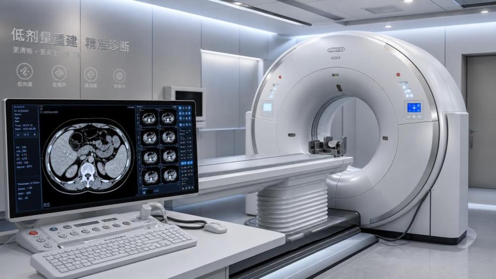
In earlier CT practice, dose reduction often meant adjusting tube current, voltage, pitch, and scan length.
Today, medical imaging reconstruction technology changes the equation by recovering useful signal from noisier or incomplete acquisition data.
This shift matters because modern clinical imaging must satisfy safety, throughput, reimbursement, and diagnostic confidence simultaneously.
Hospitals cannot simply reduce dose if lesions become less visible or artifacts distort anatomy.
Therefore, reconstruction quality is now judged against task-based performance, not only visual smoothness.
For AMDS, this trend connects physics, clinical compliance, and health economics into one measurable technology question.
Why Medical Imaging Reconstruction Technology Can Lower Exposure
Dose reduction is possible because imaging systems collect signals that are noisy, incomplete, or distorted by patient and hardware factors.
Reconstruction algorithms estimate the most clinically plausible image from this imperfect data.
The most important trend is the move from filtered back projection toward iterative and AI-assisted methods.
Medical imaging reconstruction technology cuts dose best when acquisition design and reconstruction design are validated together.
A low-dose protocol is not automatically safe because an algorithm produces a clean-looking image.
The Real Test Is Diagnostic Confidence, Not Cosmetic Smoothness
A visually pleasing image can still fail if microcalcifications, small nodules, stents, or low-contrast lesions become less detectable.
This is why medical imaging reconstruction technology must be evaluated using clinical tasks and observer studies.
Key indicators include contrast-to-noise ratio, modulation transfer function, noise power spectrum, and lesion detectability.
However, numbers alone are not enough for high-risk clinical use.
Radiology performance depends on anatomy, disease prevalence, patient size, scanner generation, and reporting workflow.
For example, lung screening benefits from lower-dose CT because air-tissue contrast is naturally high.
Abdominal oncology imaging is harder because low-contrast liver lesions demand stronger signal integrity.
Therefore, medical imaging reconstruction technology should be judged by indication-specific evidence.
Dose Cutting Is Strongest In Repeated Imaging Pathways
The largest cumulative benefit appears in populations requiring repeated scans.
- Cancer surveillance, where multiple CT scans follow treatment response.
- Pediatric imaging, where lifetime radiation sensitivity is higher.
- Cardiac CT, where motion and timing affect repeat probability.
- Emergency imaging, where speed and diagnostic clarity must coexist.
In these settings, medical imaging reconstruction technology can reduce cumulative exposure while protecting clinical utility.
CT Leads The Dose Conversation, But MRI Also Benefits
Dose reduction is most directly associated with CT because ionizing radiation is involved.
Yet medical imaging reconstruction technology also changes MRI, where the comparable pressure is scan time and motion failure.
Compressed sensing, parallel imaging, and AI reconstruction can shorten MRI exams while preserving diagnostic contrast.
Shorter MRI slots reduce sedation demand, motion artifacts, and scheduling delays.
This creates a safety impact even without radiation dose.
Photon-counting CT adds another layer by improving energy discrimination and spatial resolution.
When paired with advanced reconstruction, it may lower contrast dose, radiation dose, or both in selected exams.
The emerging direction is not one universal low-dose setting.
It is patient-adaptive, anatomy-specific, and task-aware imaging.
Regulation Is Turning Reconstruction Claims Into Audit Questions
Claims about dose reduction now face stronger scrutiny under clinical safety and market access frameworks.
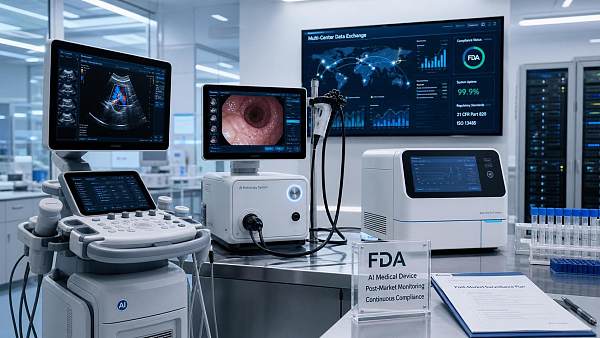
A reconstruction feature must prove repeatable performance, cybersecurity protection, software lifecycle control, and post-market monitoring.
This is especially important when medical imaging reconstruction technology uses AI models updated over time.
Version changes can alter image texture, lesion conspicuity, and radiologist perception.
Regulatory evidence therefore needs traceability from raw acquisition through reconstructed output.
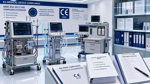
CE MDR and FDA expectations increasingly favor transparent risk management and clinically meaningful performance endpoints.
The question is no longer whether medical imaging reconstruction technology can improve images.
The harder question is whether improvement remains stable under real clinical variability.
Business Impact Extends Across Imaging, IVD, ICU, And Operating Rooms
Although reconstruction is an imaging topic, its impact touches the broader digital healthcare ecosystem.
Better imaging can reduce unnecessary biopsies, support IVD correlation, and guide minimally invasive treatment planning.
In critical care, faster and safer imaging supports decisions for ventilation, ECMO, and emergency interventions.
In operating rooms, accurate preoperative imaging improves navigation, positioning, and endoscopic planning.
This is why medical imaging reconstruction technology is becoming part of enterprise clinical value, not only scanner specification.
Under DRG payment pressure, dose reduction alone may not justify investment.
The stronger case combines fewer repeats, shorter slots, higher confidence, better triage, and reduced downstream ambiguity.
What Should Be Watched Before Trusting A Low-Dose Claim
Medical imaging reconstruction technology should be assessed through a structured lens before protocol adoption or capital decisions.
- Check whether validation includes real patient data, not only phantom measurements.
- Compare low-dose images with standard-dose references for the same clinical task.
- Evaluate whether image texture remains familiar enough for consistent reporting.
- Track repeat scan rates before and after reconstruction deployment.
- Confirm compatibility with dose monitoring and reporting systems.
- Review how software updates are documented and clinically communicated.
A strong low-dose program treats reconstruction as part of a controlled imaging pathway.
It does not treat the algorithm as a black-box shortcut.
The Next Phase Will Be Task-Aware And Patient-Adaptive
The next wave of medical imaging reconstruction technology will likely become more personalized and more auditable.
Algorithms will adapt to body habitus, anatomy, contrast phase, motion risk, and diagnostic question.
Protocol selection may increasingly combine scanner telemetry, prior imaging, laboratory signals, and clinical indication.
This will require stronger governance because adaptive systems can create hidden variability.
The safest direction is transparent automation with measurable clinical guardrails.
Medical imaging reconstruction technology can cut dose, but only when evidence, workflow, and compliance move together.
The winning standard is not the lowest possible dose.
It is the lowest justified dose that still preserves reliable, timely, and clinically actionable diagnosis.
Actionable Direction For Safer Imaging Transformation
Organizations evaluating reconstruction performance should begin with high-volume, high-repeat, or radiation-sensitive pathways.
Build a baseline using current dose indices, repeat rates, report confidence, and turnaround times.
Then test medical imaging reconstruction technology against specific clinical questions rather than generic image quality claims.
AMDS follows this evidence-first view across imaging, IVD, life support, operating rooms, and endoscopic systems.
The next practical step is to map reconstruction claims to safety metrics, regulatory evidence, and economic outcomes.
That is how dose reduction becomes a defensible clinical strategy, not merely a software promise.
Recommended News
 Aug 28, 2026Planning Operating Room Technology for a High-Throughput Surgery CenterOperating room technology for a surgery center: plan workflows, integration, uptime, and safety to boost throughput and create future-ready surgical rooms.
 Aug 27, 2026Building Medical Compliance Analytics for Middle East Market AccessMedical compliance analytics Middle East: build device-specific evidence maps, align local requirements, and accelerate market access with confidence.
 Aug 26, 2026How Surgical Equipment Digitalization Is Reshaping Hospital Investment in Latin AmericaSurgical equipment digitalization Latin America is reshaping hospital investment, from smart ORs to ROI-driven upgrades. Explore what buyers now value most.
 Aug 25, 2026How to Evaluate a Medical Compliance Intelligence Provider for Global Market AccessMedical compliance intelligence provider selection can make or break global market access. Learn how to assess technical depth, update quality, and commercial fit before choosing.
 Aug 24, 2026How Advanced Clinical Imaging Is Improving Cardiology Diagnosis and Treatment PlanningAdvanced clinical imaging cardiology is transforming diagnosis and treatment planning with clearer anatomy, tissue insight, and procedural guidance. Discover how it reduces uncertainty and supports smarter cardiac decisions.
 Aug 23, 2026How to Source Medical Devices in North America: Supplier Vetting and Market Access RisksMedical device sourcing North America made practical: learn how to vet suppliers, reduce compliance risks, and protect market access with smarter procurement decisions.
 Aug 22, 2026What Are Integrated Critical Care Systems and How Do Hospitals Evaluate Them?Critical care systems integrated: learn how hospitals assess ICU platforms for interoperability, alarm safety, workflow efficiency, and long-term value before buying.
 Aug 21, 2026How to Evaluate High-Resolution Diagnostic Imaging Systems for Detail-Critical ExamsDiagnostic imaging systems high resolution: learn how to compare detail-critical performance, workflow fit, dose efficiency, and ROI to choose the right system with confidence.
 Aug 20, 2026How to Evaluate Medical Equipment Performance Before Purchase?Medical equipment evaluation performance starts before purchase. Learn how to assess safety, workflow fit, compliance, uptime, and total cost to choose devices that truly perform.
 Aug 19, 2026How to Evaluate Small-Footprint Laboratory Diagnostic Equipment for Space-Limited LabsLaboratory diagnostic equipment small footprint choices require more than size checks. Learn how to assess workflow, throughput, compliance, and service access for smarter lab buying.
 Aug 18, 2026Biochemical Molecular Diagnostics Explained: Methods, Clinical Uses, and Lab Selection CriteriaBiochemical molecular diagnostics explained: compare key methods, clinical applications, and lab selection criteria to choose reliable, compliant testing with confidence.
 Aug 17, 2026How to Choose Diagnostic Equipment Solutions for a Clinic: Key Factors That Affect Workflow and CostDiagnostic equipment solutions clinic buyers can use to improve workflow, control total cost, and avoid hidden service risks. Learn the key factors before you invest.
 Aug 16, 2026How to Choose a Blood Cell Analyzer for a Small Lab: Throughput, CBC Menu, and CostBlood cell analyzers small lab buyers can compare throughput, CBC menu, and total operating cost with confidence. Learn how to avoid waste, reduce downtime, and choose the right fit.
 Aug 16, 2026FDA Approval of AMDS Hybrid Prosthesis Removes IRB Step for Faster U.S. AccessFDA approval of AMDS Hybrid Prosthesis removes the IRB step, speeding U.S. access for acute aortic dissection care and reshaping hospital procurement and supporting equipment demand.
 Aug 15, 2026What Hospital Technology Infrastructure Is Needed for Reliable Interoperability?Hospital technology infrastructure interoperability depends on secure networks, integration engines, identity services, and governance. Discover what hospitals need for reliable, scalable connectivity.
 Aug 14, 2026What to Look for in an Advanced Diagnostic Systems Distributor Before You BuyAdvanced diagnostic systems distributor: learn what to check before buying, from compliance and technical depth to delivery reliability and after-sales support. Make a smarter procurement decision today.
 Aug 14, 2026China Temporarily Bans Helium ExportsChina Temporarily Bans Helium Exports: learn how the July 10, 2026 rule may disrupt MRI, ECMO, and anesthesia device supply chains, compliance, and delivery planning for global buyers.
 Aug 13, 2026How CT Reconstruction Technology Impacts Image Quality, Dose, and WorkflowMedical imaging reconstruction technology CT shapes image quality, dose, and workflow. Learn how smarter CT reconstruction improves diagnostics and efficiency.
 Aug 13, 2026FDA clears AMDS PMA, easing U.S. hospital accessFDA clears AMDS PMA, easing U.S. hospital access for Artivion’s aortic arch device and speeding procurement, compliance, and deployment.
 Aug 12, 2026Hospital Diagnostic Equipment Comparison: What Buyers Should Evaluate Beyond PriceDiagnostic equipment comparison hospital buyers can trust: learn how to evaluate clinical performance, uptime, compliance, integration, and long-term ROI beyond price.
 Aug 12, 2026FDA Draft Guidance Reshapes AI Device ComplianceFDA Draft Guidance reshapes AI device compliance with new postmarket monitoring expectations for AI/ML-enabled SaMD. Learn what it means for U.S. market access, distributors, and exporters.
 Aug 11, 2026How to Evaluate a Diagnostic Medical Systems Manufacturer for Quality and Market AccessDiagnostic medical systems manufacturer selection starts with more than specs. Learn how to assess quality systems, regulatory readiness, service uptime, and market access before you buy.
 Aug 11, 2026EU MDR Deadline Extended for ECMO and ICU DevicesEU MDR deadline extended for ECMO and ICU devices to 2027. Learn what Regulation (EU) 2026/1347 means for compliance, exports, procurement, and roadmap filing.
 Aug 10, 2026How to Evaluate Laboratory Automation Systems in Europe for Multi-Site Lab OperationsLaboratory automation systems Europe buyers can use this guide to compare interoperability, compliance, service coverage, and ROI for multi-site lab operations with confidence.







Related News
- 00
0000-00
Medical Imaging Diagnostics Systems: Core Modalities, Use Cases, and Trade-Offs - 00
0000-00
AI in Medical Imaging Radiology: Where It Improves Reading Speed and Where It Falls Short - 00
0000-00
Saudi SFDA Opens 18-Day Fast Track for DR Devices - 00
0000-00
SFDA Fast-Track Cuts DR Device Approval to 22 Days - 00
0000-00
Diagnostic Imaging Technology Comparison: DR vs CT vs MRI for Hospital Planning
Weekly Insights
Stay ahead with our curated technology reports delivered every Monday.