
Is endoscopic imaging technology worth upgrading?
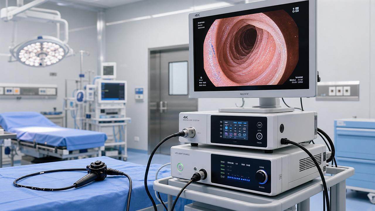




For healthcare systems, the decision to upgrade endoscopic imaging technology is no longer just about sharper visuals.
It is about clinical confidence, workflow efficiency, compliance readiness, and measurable return on investment.
As 4K, 3D visualization, AI enhancement, anti-fog optics, and ultra-thin platforms reshape minimally invasive care, upgrade decisions require structured judgment.
Is endoscopic imaging technology worth upgrading?
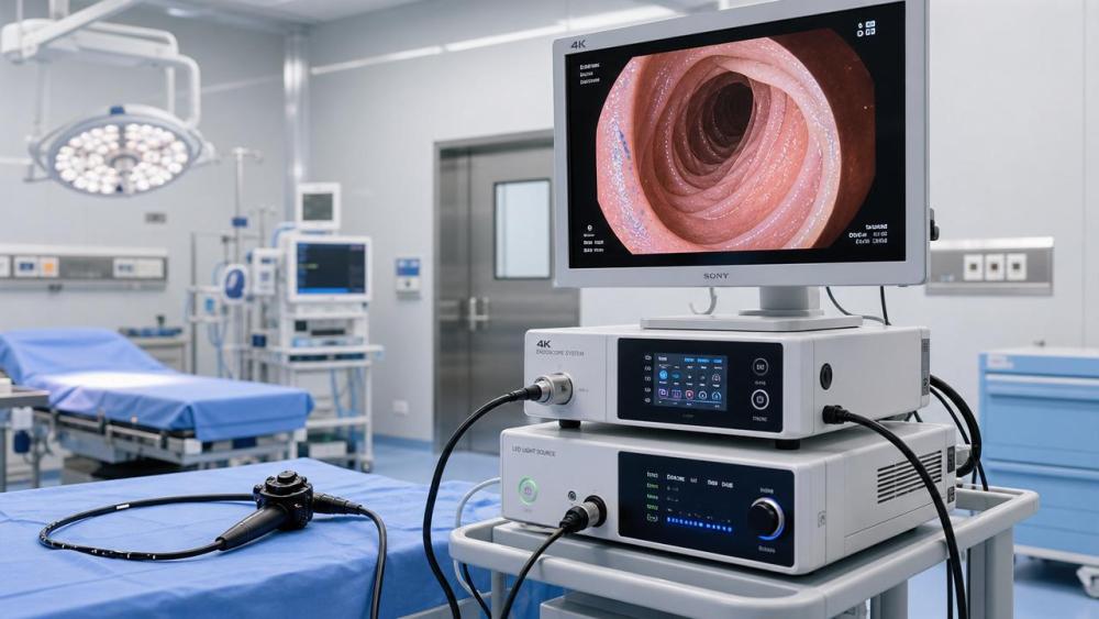
Endoscopic imaging technology is worth upgrading when it improves diagnosis, reduces procedural uncertainty, and supports safer minimally invasive interventions.
The strongest upgrade cases appear when older systems limit visibility, slow procedures, or fail to meet modern documentation standards.
A useful decision should connect image quality, clinical workflow, infection control, device interoperability, and financial performance.
The goal is not to buy the newest platform.
The goal is to prove that newer endoscopic imaging technology creates measurable value inside real operating rooms and diagnostic suites.
Why a checklist is essential before upgrading endoscopic imaging technology
Endoscopy systems now combine optics, sensors, processors, software, displays, insufflation, recording, and network connectivity.
A weakness in any layer can reduce the expected benefit of an upgrade.
Checklist-based evaluation prevents decisions driven only by demo-room brightness, marketing claims, or isolated surgeon preference.
It also helps compare vendors using evidence that matters clinically and operationally.
For AMDS, endoscopic imaging technology belongs to the critical infrastructure of minimally invasive medicine.
It should be assessed with the same rigor applied to CT, MRI, IVD, life support, and operating room systems.
Core checklist for evaluating endoscopic imaging technology
- Compare native resolution, sensor size, color reproduction, and low-light performance under realistic cavity conditions, not only vendor showroom demonstrations.
- Verify whether 4K or 3D imaging improves anatomical orientation, tissue differentiation, and confidence during high-risk dissection or diagnostic inspection.
- Test anti-fog optics, smoke resistance, lens cleaning behavior, and light stability during long procedures with frequent instrument exchanges.
- Assess AI-assisted image enhancement for edge clarity, bleeding visibility, mucosal pattern recognition, and false highlight control.
- Confirm compatibility with existing scopes, camera heads, light sources, monitors, recording systems, carts, and operating room integration platforms.
- Measure setup time, boot speed, white balance workflow, user interface logic, and cleaning turnover between consecutive procedures.
- Review cybersecurity, patient data storage, audit trails, video export formats, and hospital network access requirements before deployment.
- Calculate total ownership cost, including scopes, service contracts, consumables, sterilization impact, training, downtime, and future software upgrades.
- Request clinical evidence showing reduced procedure time, better lesion detection, fewer conversions, or improved documentation quality.
- Validate regulatory documentation, CE MDR or FDA status, service traceability, and post-market surveillance commitments from the supplier.
This checklist turns endoscopic imaging technology from a visual preference into a structured capital investment decision.
Clinical value: what better visualization must actually prove
Sharper images are valuable only when they change clinical action.
Modern endoscopic imaging technology should help detect subtle lesions, clarify tissue planes, and reduce uncertainty in narrow anatomical spaces.
In gastrointestinal procedures, enhanced visualization may support recognition of vascular patterns, mucosal texture, and lesion boundaries.
In laparoscopy, higher contrast and depth perception may improve dissection around vessels, ducts, and inflamed tissue.
In urology and gynecology, compact scopes and stable illumination can improve access in confined fields.
However, every clinical claim should be tested against case mix, operator experience, and local procedural priorities.
Use objective evaluation metrics
- Track image-related interruptions, including fogging, cleaning pauses, glare, signal loss, and repeated camera adjustments.
- Record procedure duration before and after implementation for comparable case types and similar clinical complexity.
- Review documentation quality, still-image clarity, video archiving reliability, and usability during multidisciplinary case review.
- Monitor conversion rates, repeat procedures, missed lesion concerns, and complications where visualization may be contributory.
Endoscopic imaging technology should be judged through repeatable metrics, not subjective enthusiasm alone.
Workflow and operating room efficiency considerations
A high-resolution platform can still fail if it slows daily workflow.
Endoscopic imaging technology must support quick setup, predictable image calibration, simple source switching, and fast troubleshooting.
The best systems reduce cognitive load during procedures.
They make essential controls accessible without forcing staff to navigate complex menus at critical moments.
Workflow gains also depend on cleaning, sterilization, and scope availability.
Ultra-thin flexible endoscopes may improve access, but they must withstand repeated reprocessing without frequent repair.
Operational checks before purchase
- Run trial cases across morning, emergency, and back-to-back lists to expose real setup and turnover problems.
- Ask biomedical teams to inspect cabling, connectors, spare parts, software access, and preventive maintenance requirements.
- Simulate video recording, image export, case labeling, and integration with electronic medical record workflows.
- Confirm that older equipment can remain available during phased deployment, training, and service interruptions.
These checks reveal whether endoscopic imaging technology improves throughput or merely adds another complex device layer.
Scenario guidance for different endoscopy environments
High-volume diagnostic endoscopy
In high-volume diagnostic settings, speed and consistency matter as much as peak image quality.
Endoscopic imaging technology should support rapid case documentation, stable visualization, and reliable lesion capture across many examinations.
The priority is repeatable diagnostic confidence with minimal downtime.
Advanced minimally invasive surgery
In complex laparoscopy or thoracoscopy, 3D depth perception and precise color rendering may carry stronger value.
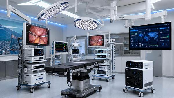
Endoscopic imaging technology should improve orientation during dissection, suturing, bleeding control, and navigation around fragile anatomy.
Here, surgeon adaptation time and ergonomics deserve careful attention.
Resource-sensitive hospitals
Where budgets are constrained, a staged upgrade may be more practical than a complete platform replacement.
Camera processors, light sources, displays, and recording modules can sometimes be upgraded in carefully planned phases.
The main requirement is a realistic compatibility map.
Commonly overlooked risks in endoscopic imaging technology upgrades
Hidden interoperability limits. New processors may not fully support older scopes, preferred monitors, or existing video routing infrastructure.
This can increase cost after approval and delay go-live timelines.
Insufficient training time. Better endoscopic imaging technology may introduce new modes, overlays, presets, and documentation tools.
Without structured training, advanced functions may remain unused or inconsistently applied.
Overdependence on image enhancement. AI enhancement and digital contrast are supportive tools, not replacements for clinical judgment.
Evaluation should include false emphasis, artifacts, and behavior under bleeding, smoke, or fluid distortion.
Unclear service response. Endoscopic imaging technology is mission-critical during scheduled and emergency procedures.
Service agreements should specify response time, loaner availability, software support, and repair cost transparency.
Weak data governance. Networked endoscopy platforms may store images, videos, identifiers, and procedural metadata.
Cybersecurity review should occur before purchase, not after installation.
ROI checklist: when the upgrade becomes financially defensible
Return on investment is not limited to acquisition price.
Endoscopic imaging technology can influence case throughput, repeat procedure rates, training efficiency, equipment downtime, and documentation quality.
- Estimate annual procedure volume by specialty, then identify cases where visualization directly affects time, confidence, or complication risk.
- Compare maintenance cost trends from aging systems against projected service cost for the proposed new platform.
- Quantify avoidable downtime, including canceled cases, delayed starts, emergency rentals, and staff waiting time.
- Include documentation value, especially for tumor boards, quality audits, teaching, claims review, and medicolegal records.
- Model phased deployment to reduce capital shock while preserving compatibility with existing clinical workflows.
Under DRG-style payment pressure, ROI must connect equipment spending to efficiency, safety, and resource predictability.
Practical implementation plan
Start with a baseline audit of current systems.
List image complaints, repair frequency, procedure delays, unsupported formats, and clinical gaps reported during routine use.
Next, define required outcomes before reviewing vendor proposals.
Examples include better lesion documentation, reduced fogging interruptions, improved 3D navigation, or faster digital archiving.
Then perform controlled trials using comparable cases and agreed scoring sheets.
Avoid judging endoscopic imaging technology from one impressive procedure or one enthusiastic demonstration.
Finally, negotiate implementation support, training, warranty terms, cybersecurity documentation, and service escalation before signing.
Minimum decision file
- Current-state audit with repair history, age profile, compatibility gaps, and recurring clinical complaints.
- Trial evaluation records covering image quality, workflow impact, user experience, and documentation performance.
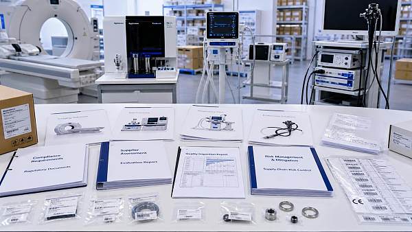
- Regulatory and compliance documents, including market authorization, cybersecurity statements, and post-market support commitments.
- Financial model showing total ownership cost, phased options, service exposure, and expected operational gains.
Conclusion and next action
Endoscopic imaging technology is worth upgrading when it delivers more than visual improvement.
A strong upgrade improves diagnostic certainty, surgical control, workflow reliability, compliance readiness, and economic predictability.
The safest path is to evaluate endoscopic imaging technology through evidence, scenario testing, and total ownership analysis.
Begin with a structured audit, define clinical success metrics, run controlled trials, and compare vendors against measurable requirements.
When the data supports better care and better operations, upgrading endoscopic imaging technology becomes a strategic clinical investment.
Recommended News
 Aug 29, 2026How to Select a Hematology Analyzer With Reliable Reticulocyte CountingHematology analyzers retic selection guide: compare precision, flags, workflow, QC, and connectivity to achieve reliable reticulocyte counting.
 Aug 28, 2026Planning Operating Room Technology for a High-Throughput Surgery CenterOperating room technology for a surgery center: plan workflows, integration, uptime, and safety to boost throughput and create future-ready surgical rooms.
 Aug 27, 2026Building Medical Compliance Analytics for Middle East Market AccessMedical compliance analytics Middle East: build device-specific evidence maps, align local requirements, and accelerate market access with confidence.
 Aug 26, 2026How Surgical Equipment Digitalization Is Reshaping Hospital Investment in Latin AmericaSurgical equipment digitalization Latin America is reshaping hospital investment, from smart ORs to ROI-driven upgrades. Explore what buyers now value most.
 Aug 25, 2026How to Evaluate a Medical Compliance Intelligence Provider for Global Market AccessMedical compliance intelligence provider selection can make or break global market access. Learn how to assess technical depth, update quality, and commercial fit before choosing.
 Aug 24, 2026How Advanced Clinical Imaging Is Improving Cardiology Diagnosis and Treatment PlanningAdvanced clinical imaging cardiology is transforming diagnosis and treatment planning with clearer anatomy, tissue insight, and procedural guidance. Discover how it reduces uncertainty and supports smarter cardiac decisions.
 Aug 23, 2026How to Source Medical Devices in North America: Supplier Vetting and Market Access RisksMedical device sourcing North America made practical: learn how to vet suppliers, reduce compliance risks, and protect market access with smarter procurement decisions.
 Aug 22, 2026What Are Integrated Critical Care Systems and How Do Hospitals Evaluate Them?Critical care systems integrated: learn how hospitals assess ICU platforms for interoperability, alarm safety, workflow efficiency, and long-term value before buying.
 Aug 21, 2026How to Evaluate High-Resolution Diagnostic Imaging Systems for Detail-Critical ExamsDiagnostic imaging systems high resolution: learn how to compare detail-critical performance, workflow fit, dose efficiency, and ROI to choose the right system with confidence.
 Aug 20, 2026How to Evaluate Medical Equipment Performance Before Purchase?Medical equipment evaluation performance starts before purchase. Learn how to assess safety, workflow fit, compliance, uptime, and total cost to choose devices that truly perform.
 Aug 19, 2026How to Evaluate Small-Footprint Laboratory Diagnostic Equipment for Space-Limited LabsLaboratory diagnostic equipment small footprint choices require more than size checks. Learn how to assess workflow, throughput, compliance, and service access for smarter lab buying.
 Aug 18, 2026Biochemical Molecular Diagnostics Explained: Methods, Clinical Uses, and Lab Selection CriteriaBiochemical molecular diagnostics explained: compare key methods, clinical applications, and lab selection criteria to choose reliable, compliant testing with confidence.
 Aug 17, 2026How to Choose Diagnostic Equipment Solutions for a Clinic: Key Factors That Affect Workflow and CostDiagnostic equipment solutions clinic buyers can use to improve workflow, control total cost, and avoid hidden service risks. Learn the key factors before you invest.
 Aug 16, 2026How to Choose a Blood Cell Analyzer for a Small Lab: Throughput, CBC Menu, and CostBlood cell analyzers small lab buyers can compare throughput, CBC menu, and total operating cost with confidence. Learn how to avoid waste, reduce downtime, and choose the right fit.
 Aug 16, 2026FDA Approval of AMDS Hybrid Prosthesis Removes IRB Step for Faster U.S. AccessFDA approval of AMDS Hybrid Prosthesis removes the IRB step, speeding U.S. access for acute aortic dissection care and reshaping hospital procurement and supporting equipment demand.
 Aug 15, 2026What Hospital Technology Infrastructure Is Needed for Reliable Interoperability?Hospital technology infrastructure interoperability depends on secure networks, integration engines, identity services, and governance. Discover what hospitals need for reliable, scalable connectivity.
 Aug 14, 2026What to Look for in an Advanced Diagnostic Systems Distributor Before You BuyAdvanced diagnostic systems distributor: learn what to check before buying, from compliance and technical depth to delivery reliability and after-sales support. Make a smarter procurement decision today.
 Aug 14, 2026China Temporarily Bans Helium ExportsChina Temporarily Bans Helium Exports: learn how the July 10, 2026 rule may disrupt MRI, ECMO, and anesthesia device supply chains, compliance, and delivery planning for global buyers.
 Aug 13, 2026How CT Reconstruction Technology Impacts Image Quality, Dose, and WorkflowMedical imaging reconstruction technology CT shapes image quality, dose, and workflow. Learn how smarter CT reconstruction improves diagnostics and efficiency.
 Aug 13, 2026FDA clears AMDS PMA, easing U.S. hospital accessFDA clears AMDS PMA, easing U.S. hospital access for Artivion’s aortic arch device and speeding procurement, compliance, and deployment.
 Aug 12, 2026Hospital Diagnostic Equipment Comparison: What Buyers Should Evaluate Beyond PriceDiagnostic equipment comparison hospital buyers can trust: learn how to evaluate clinical performance, uptime, compliance, integration, and long-term ROI beyond price.
 Aug 12, 2026FDA Draft Guidance Reshapes AI Device ComplianceFDA Draft Guidance reshapes AI device compliance with new postmarket monitoring expectations for AI/ML-enabled SaMD. Learn what it means for U.S. market access, distributors, and exporters.
 Aug 11, 2026How to Evaluate a Diagnostic Medical Systems Manufacturer for Quality and Market AccessDiagnostic medical systems manufacturer selection starts with more than specs. Learn how to assess quality systems, regulatory readiness, service uptime, and market access before you buy.
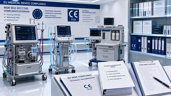
 Aug 11, 2026EU MDR Deadline Extended for ECMO and ICU DevicesEU MDR deadline extended for ECMO and ICU devices to 2027. Learn what Regulation (EU) 2026/1347 means for compliance, exports, procurement, and roadmap filing.

Related News
- 00
0000-00
EU MDR Transition Ends, Endoscope Re-Certification Becomes Urgent - 00
0000-00
EU MDR Annex XVI Now Requires Separate CE for Accessories - 00
0000-00
EU MDR Transition Ends for Endoscopy Device Imports - 00
0000-00
EU MDR Rule Triggers CER Updates for Endoscopy Devices - 00
0000-00
EU MDR Update Sets 12-Month CER Rule for Ultrasound and Endoscopy
Weekly Insights
Stay ahead with our curated technology reports delivered every Monday.